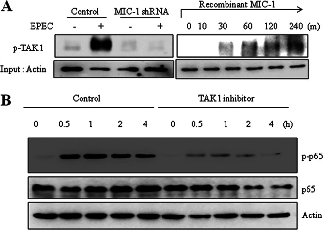
Fig 6.

Involvement of TAK1 in MIC-1-linked signaling in EPEC-infected intestinal epithelial cells. (A) Stable cell lines (control and HCT-8 cells expressing MIC-1-specific shRNA) were infected with EPEC at a ratio of 50:1 (bacteria to cells) for 4 h. Phosphorylated TAK1 (p-TAK1) was detected by Western blotting (left). HCT-8 cells were treated with 10 ng/ml recombinant MIC-1 for the indicated times (right). Total proteins from the epithelial cellular lysate were subjected to Western blot analysis. (B) HCT-8 cells were pretreated with vehicle (dimethyl sulfoxide) or 100 nM (5Z)-7-oxozeaenol (5Z-OXO), a TAK1 inhibitor, and then infected with EPEC for the indicated times. Total proteins from the epithelial cellular lysate were subjected to Western blot analysis. All results are representative of three independent experiments.
