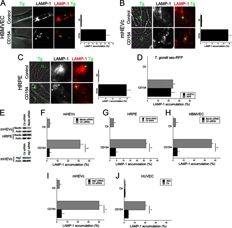
Fig 3.
CD40 induces vacuole-lysosome fusion in T. gondii-infected nonhematopoietic cells, dependent on the autophagy machinery. (A to C) HBMVEC (A), hmCD40-mHEVc (B), and hCD40-HRPE (C) were incubated with or without CD154 and infected with RH T. gondii-YFP. At 8 h postinfection, LAMP-1 expression was assessed by immunofluorescence, and the percentages of cells that exhibited accumulation of LAMP-1 around the parasites (arrowheads) were quantitated. (D) hmCD40-mHEVc were stimulated with or without CD154 and infected with T. gondii expressing Sec-RFP. Percentages of cells that exhibited accumulation of LAMP-1 around vacuoles that associate with RFP (parasitophorous vacuoles) were quantitated. (E) hmCD40-mHEVc or hCD40-HRPE were transfected with control siRNA, Beclin 1 siRNA, or Atg7 siRNA. Expression of Beclin 1, Atg7, and actin was assessed by immunoblotting. (F to H) hmCD40-mHEVc (F), hCD40-HRPE (G), or HBMVEC (H) transfected with control or Beclin 1 siRNA were incubated with or without CD154 and infected with T. gondii expressing YFP. Cells were immunostained for LAMP-1 and analyzed by fluorescence microscopy for accumulation of LAMP-1 around the parasite. (I) hmCD40-mHEVc were transfected with either control siRNA or Atg7 siRNA. Cells were incubated with or without CD154 and infected with T. gondii expressing YFP. Cells were immunostained for LAMP-1 expression and then analyzed by fluorescence microscopy for accumulation of LAMP-1 around the parasite. (J) HUVEC were incubated with or without CD154, infected with T. gondii-YFP, and treated with or without 3-MA. LAMP-1 expression was assessed by immunofluorescence. Results are shown as means ± SEM and are representative of 3 independent experiments. **, P ≤ 0.01; ***, P ≤ 0.001.

