
Fig 3.
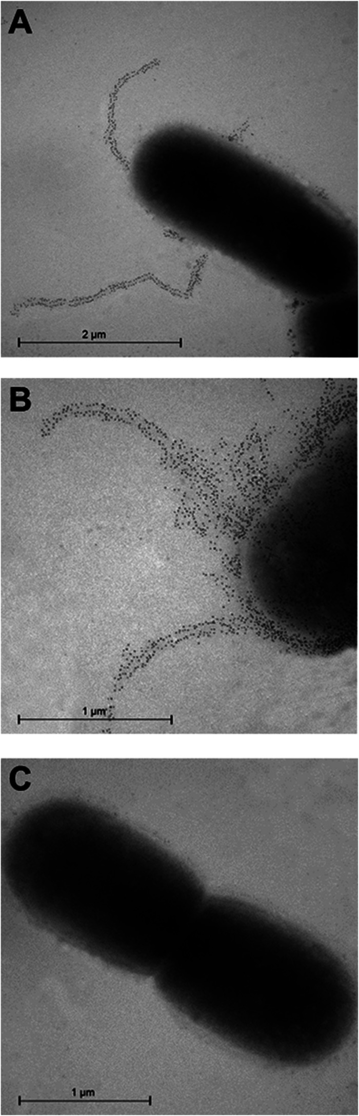
Immunogold electron microscopy with anti-FlfA immune serum. G. anatis 12656-12 WT cells (A and B) or ΔflfA mutant cells (C) were incubated with anti-FlfA immune serum and labeled with a secondary antibody conjugated to 10-nm gold particles. Two fimbrial cell populations were observed: a low-level population (A) and a hyperfimbriated population (B).