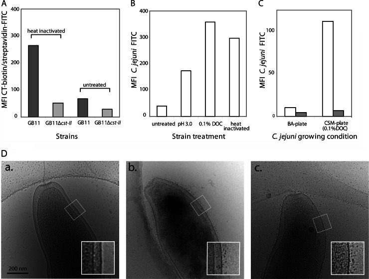
Fig 2.
Exposure of ganglioside mimics on the surface of C. jejuni. (A) Flow-cytometric analysis of the expression of ganglioside mimics on the surface of C. jejuni. Bacteria were either heat inactivated or left untreated and then subsequently incubated with CT-biotin and streptavidin-FITC. (B) Flow-cytometric analysis of the binding of FITC-labeled C. jejuni strain GB11 to THP-1-Sn cells. Bacteria grown on BA plates were left untreated, incubated for 1 h in PBS (pH 3.0) or in PBS (pH 7.0) containing 0.1% DOC, or heat inactivated. (C) Flow-cytometric analysis of the binding of FITC-labeled C. jejuni strain GB11 to THP-1-Sn cells. Bacteria were grown on either BA plates or CSM plates. THP-1-Sn cells were left untreated (white bars) or were pretreated to block Sn binding using an anti-hSn antibody (gray bars). (D) Cryo-EM visualization of C. jejuni strain GB11 grown on BA plates and left untreated (a), grown on BA plates and heat inactivated (b), or grown on CSM plates and left untreated (c). The bacteria were incubated with CT-biotin followed by streptavidin-conjugated quantum dots.

