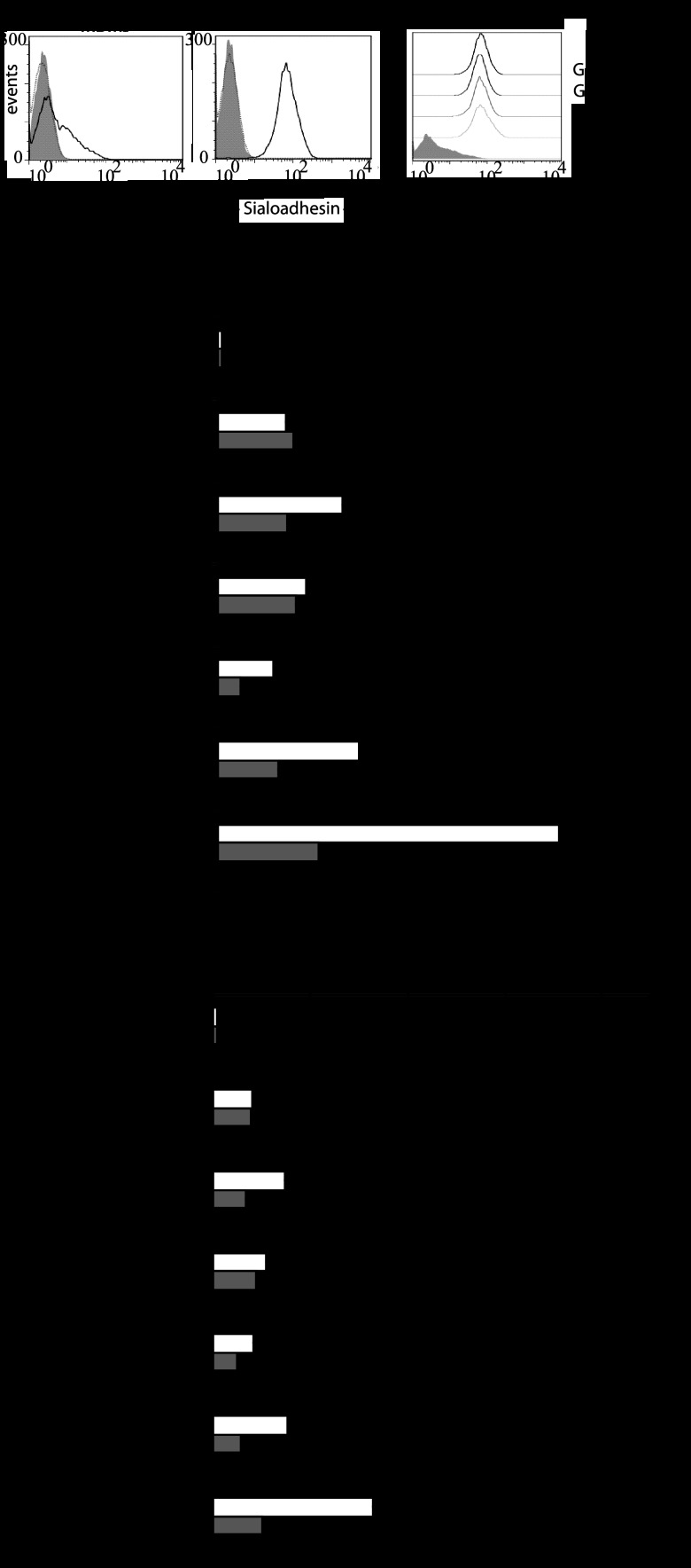
Fig 3.
Sn-specific binding of sialylated C. jejuni strains results in enhanced uptake by Sn+MDMs. Human monocyte-derived macrophages were either left untreated (MDMs) or treated with IFN-α to induce expression of Sn (Sn+MDMs), and then Sn expression and the capacity to bind and internalize C. jejuni were quantified. FITC-labeled, heat-inactivated C. jejuni stains were incubated with the cells for 2 h prior to flow-cytometric analysis. Living cells were gated and used for analysis. (A) Flow-cytometric analysis of the expression of Sn on human MDMs and Sn+MDMs in unstained cells (filled gray curve) and cells stained with anti-hSn-PE (solid-line curve) or a PE-labeled isotype control antibody (dotted-line curve). (B) Flow-cytometric analysis of the expression of Sn on untreated human MDMs or MDMs incubated for 48 h with either IFN-α (500 U/ml), E. coli LPS (10 ng/ml), C. jejuni GB11, or GB11Δcst-II LOS (10 ng/ml) and subsequently stained with anti-hSn-PE. (C) Interaction of FITC-labeled C. jejuni strains containing known ganglioside-mimicking structures with Sn+MDMs which had been untreated (white bars) or pretreated with an antibody against hSn (gray bars) or with an isotype control antibody (black bars). Results represent data from one experiment that was repeated at least one time. Means ± SD from triplicate measurements are shown. (D) Internalization of C. jejuni strains by Sn+MDMs. Sn+MDMs were incubated with FITC-labeled bacteria as described for panel C and then treated with trypan blue prior to flow-cytometric analysis to discriminate between external and internalized bacteria. For panels C and D, *, P < 0.05 (one-way ANOVA) for comparison of untreated (white bar) and isotype control treatment (black bar) versus anti-hSn treatment (gray bar). **, P < 0.05 (t test) for GB11 versus GB11Δcst-II (the combined MFI values of the untreated [white bar] and isotype control-treated [black bar] conditions were compared).

