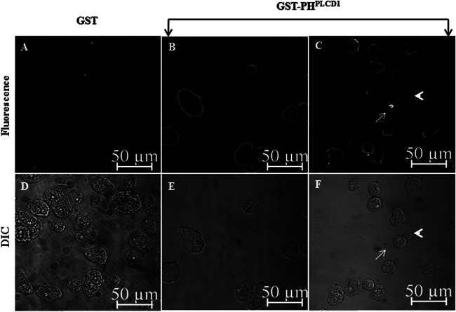
Fig 4.
The PI(4,5)P2 biosensor GST-PHPLCD1 decorates the plasma membrane of E. histolytica. Trophozoites were stained with GST or GST-PHPLCD1 and fluorescent anti-GST antibody, and immunofluorescence confocal microscopy was performed. (A) Cells stained with the control, GST, exhibited minimal staining. (B) GST-PHPLCD1 uniformly decorated the plasma membrane of the trophozoites, giving the cells a ring-like appearance. (C) In some instances, there was an enrichment of GST-PHPLCD1staining on one edge of the cell (arrow), opposite an apparent pseudopod (arrowhead). (D to F) The differential interference contrast (DIC) images corresponding to the images in panels A to C, respectively, are shown.

