Fig 5.

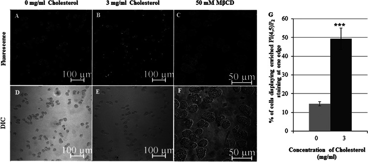
Treatment with cholesterol enhances the number of cells displaying PI(4,5)P2 enrichment at the edge opposite the forming pseudopod. Trophozoites were serum starved and exposed to either 0 mg/ml cholesterol (control), 3 mg/ml cholesterol, or the lipid raft-disrupting agent MβCD. Cells were stained with GST-PHPLCD1 and fluorescent anti-GST antibody, and immunofluorescence confocal microscopy was performed. (A and B) Cholesterol-treated cells displayed an increased PI(4,5)P2 localization on one edge of the cell compared to untreated control cells. (C) In MβCD-treated cells, PI(4,5)P2 staining was no longer confined to the plasma membrane. (D to F) The DIC images corresponding to the images in panels A to C, respectively, are shown. (G) The percentage of apparently polarized cells in control cells or after cholesterol treatment was quantified by visually scoring cells at ×20 magnification. There was a 3.4-fold increase in the number of cells displaying PI(4,5)P2 localization on one edge after cholesterol treatment compared to the number for untreated control cells. Data are the means ± SDs of 3 independent experiments (***, P < 0.001).
