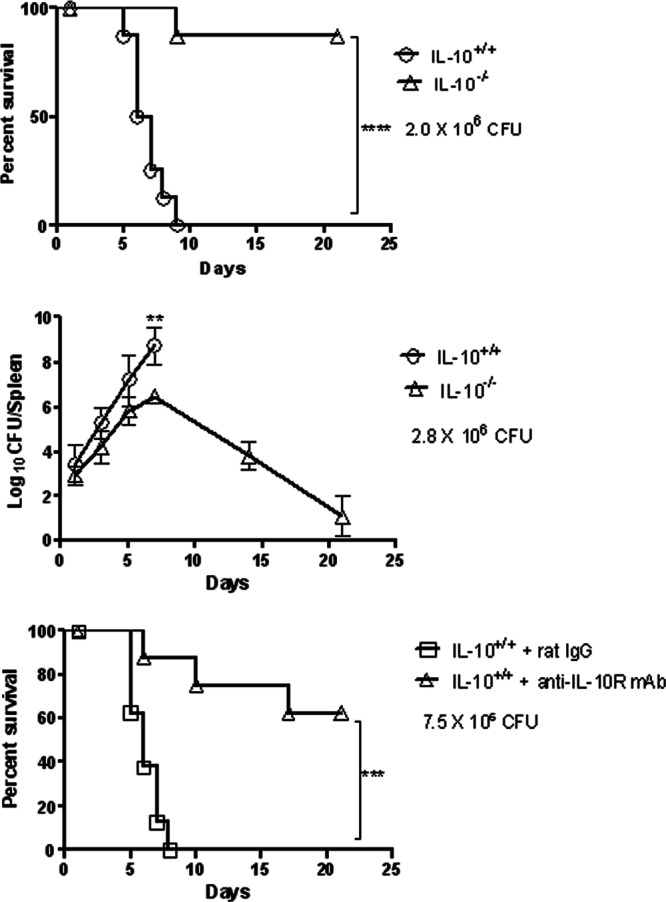
Fig 2.

Susceptibility of IL-10+/+ and IL-10−/− mice to cutaneous LVS infection. C57BL/6 mice were inoculated i.d. with 2.0 × 10⁶ CFU of LVS/mouse (top panel), 2.8 × 10⁶ CFU of LVS/mouse (middle panel), or 7.5 × 10⁶ CFU of LVS/mouse (bottom panel). In the bottom panel, the mice were also injected i.p. with 200 μg of rat IgG or neutralizing anti-IL-10R MAb on days 0, 1, 3, and 5 after bacterial challenge. In the top and bottom panels, survival was monitored daily (8 mice/group). The results are representative of two independent experiments. For animals tested for the middle panel, three mice/group were sacrificed on days 1, 3, 5, 7, 14, and 21 after infection, and their spleens were analyzed for bacterial burdens. **, P < 0.01; ***, P < 0.001; ****, P < 0.0001.
