
Fig 2.
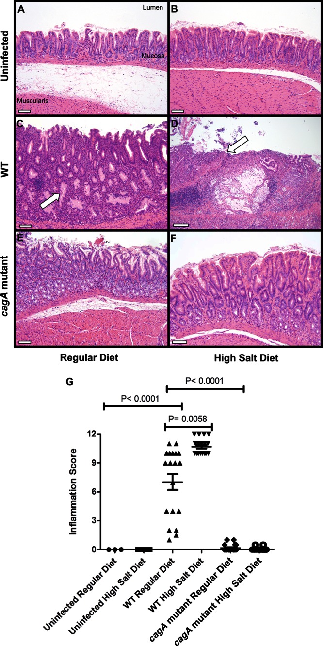
Analysis of gastric inflammation. Gerbils were infected with WT H. pylori or an isogenic cagA mutant strain and maintained on either a regular diet or a high-salt diet. As controls, uninfected gerbils were maintained on either a regular diet or a high-salt diet. At 16 weeks postinfection, gastric tissue was collected and sections were stained with hematoxylin and eosin. (A to F) Representative sections of the gastric antrum are shown. Panel C demonstrates dysplasia (arrow), and panel D demonstrates ulceration (arrow). Magnification bars indicate 100 μm. (G) Representative micrographs of gastric tissue were scored for total inflammation on a scale of 0 to 12 (sum of chronic [0 to 3] and acute [0 to 3] inflammation in both the corpus and antrum of the gerbil stomach). Animals infected with WT H. pylori and maintained on a high-salt diet had significantly higher inflammation scores than WT-infected animals maintained on a regular diet (P = 0.0058). In addition, WT-infected animals maintained on a regular diet had significantly higher inflammation scores than uninfected animals or cagA mutant-infected animals maintained on a regular diet (P < 0.0001 and P < 0.0001). Horizontal bars indicate mean total inflammation ± SEM. Statistical analyses were performed using the Mann-Whitney U-test.