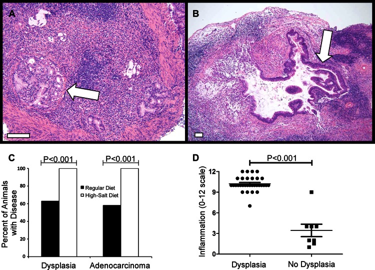
Fig 3.
Analysis of gastric dysplasia and adenocarcinoma. Gerbils were infected with WT H. pylori or an isogenic cagA mutant strain and maintained on either a regular diet or a high-salt diet. As controls, uninfected gerbils were maintained on either a regular diet or a high-salt diet. Animals were euthanized at 16 weeks postinfection. (A and B) Representative micrographs of gastric tissue derived from WT-infected animals maintained on a high-salt diet (arrows indicate tumors). Magnification bars indicate 100 μm. (C) Micrographs were evaluated for dysplastic lesions and invasive adenocarcinoma. Only animals infected with the WT strain exhibited these abnormalities. Bars indicate the percentage of animals within each group exhibiting dysplasia or gastric adenocarcinoma. Gastric cancer and dysplasia were detected significantly more frequently in the WT-infected high-salt diet group than in WT-infected regular-diet counterparts (P < 0.001). (D) Analysis of gastric inflammation in animals with gastric dysplasia. WT-infected animals maintained on a regular diet and WT-infected animals maintained on a high-salt diet were analyzed. The total gastric inflammation score (scale of 0 to 12) was higher in WT-infected animals with gastric dysplasia than in WT-infected animals without dysplastic lesions. Horizontal bars indicate mean inflammation score ± SEM for each group. Statistical analyses were performed using Fisher's exact test (C) and Mann-Whitney U analysis (D).

