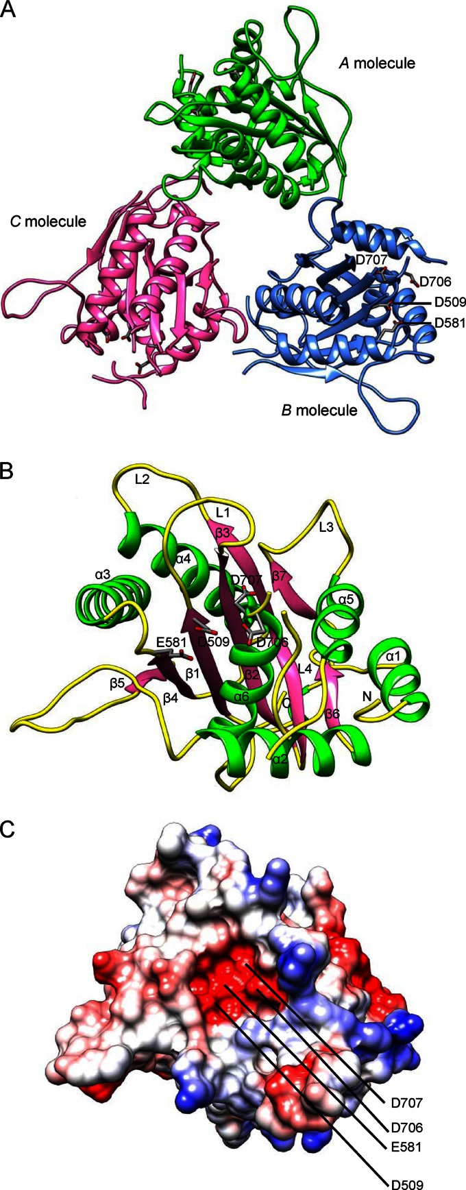
Fig 1.
The overall structure of pUL15C. (A) Ribbon representation of the pUL15C trimer viewed down the noncrystallographic 3-fold axis. A, B, and C molecules are shown in green, blue, and pink, respectively. Conserved acidic residues in the active site, D509, E581, D706, and D707, are labeled. (B) Overall structure of pUL15C in a ribbon representation. The α-helices, β-strands, and loops are in green, pink, and yellow, respectively. The loops L1, L2, L3, and L4 and the N terminus are indicated. (C) The electrostatic potential surface of pUL15C in the same view as shown in panel B. The positive potential is shown in blue, whereas the negative potential is in red.

