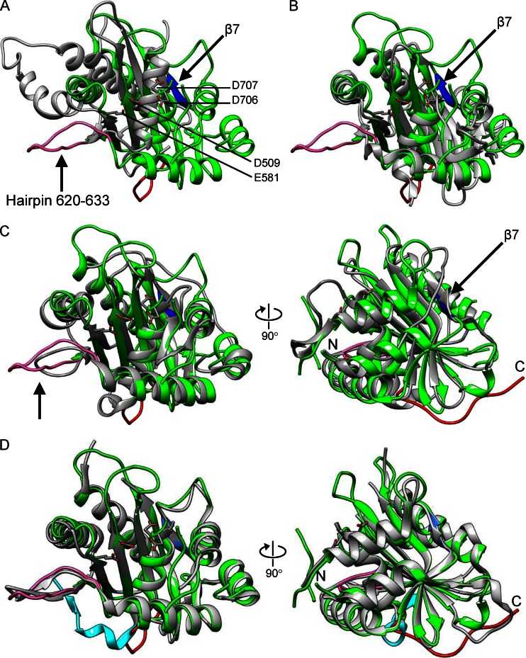
Fig 2.
Structural comparison of pUL15C with RNase H family nucleases and viral terminase nuclease domains. The pUL15C (green) is superposed with RNase H1 (A; PDB code 2QKB), phage SPP1 (B; PDB code 2WC9), phage T4 gp17 (C; PDB code 3CPE), and pUL89C (D; PDB code 3N4P). In all panels, the other protein is in gray. In panels C and D, the right views are 90° from the left ones. The pUL15C active-site residues are shown as stick models for side chains and are labeled in panel A. The β7 of pUL15C is shown in blue and the hairpin 620 to 633 is shown in pink, both of which are indicated with an arrow. In panel D, the C-terminal region encompassing residues 720 to 732 of pUL15C and the corresponding region of pUL89C are shown in red and cyan, respectively. The amino and carboxyl termini are indicated with N and C, respectively, in panels C and D.

