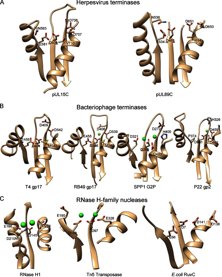
Fig 3.
Comparison of the pUL15C active-site region with those of HCMV pUL89C (A), phage terminases (B), and RNase H family nucleases (C) showing conserved features and structural variations. Conserved residues in the active sites are labeled with side chains shown as stick models. Metal ions in RNase H family nucleases are shown as green spheres. All structures are in the same view after superimposition.

