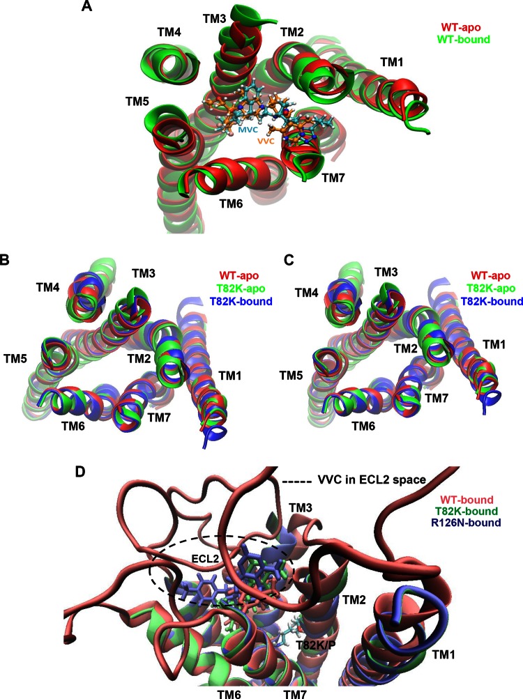
Fig 8.
TM bundle structure and orientation of the low-energy conformational state for CCR5 variants. (A) The TM helices of the unbound CCR5-WT (WT-apo) and MVC/VVC-bound CCR5-WT (WT-bound) structures predicted to have the lowest-energy conformations by the GEnSeMBLE method are superimposed. Either MVC (cyan) or VVC (orange) is docked into the TM bundle. (B and C) The TM helices of unbound CCR5-WT (WT-apo) and the unbound and VVC-bound forms of the indicated CCR5 variant are superimposed. (D) Three VVC orientations upon docking into the TM bundles are displayed in different colors, i.e., red, green, and blue, for VVC-bound CCR5-WT (WT-bound), CCR5-T82K (T82K-bound), and CCR5-R126N (R126N-bound), respectively. The extracellular loops (ECLs) of the WT-bound VVC are shown, and the space where VVC could potentially interact with ECL2 is depicted with a dotted circle. The location of Thr (T) or Lys (K) at position 82 is indicated. In all panels, the TM region at the extracellular end is facing forward.

