
Fig 1.
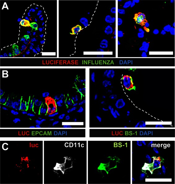
Epithelial cells and macrophages are main producers of IFN-β in lungs of mice infected with SC35M-ΔNS1. Infected global reporter mice were sacrificed at 24 h postinfection, and lung slices were simultaneously stained for luciferase and either viral antigen (A) or cellular markers (B and C). (A) Luciferase-producing cells typically expressed viral antigen and were found in (left panel) or on top of (middle panel) the epithelium of bronchi and bronchioles. A few luciferase-positive cells were also found in the alveoli (right panel). (B) Luciferase-positive cells were either epithelial cells expressing EpCAM (left panel) or lectin BS-1-positive lung macrophages (right panel). (C) Luciferase-positive cells which expressed CD11c usually also reacted with BS-1, a lectin that can specifically bind to lung macrophages. Dashed lines indicate the apical surface of the lung epithelium. Staining of nuclei (blue) was achieved with DAPI. Size bars, 20 μm.