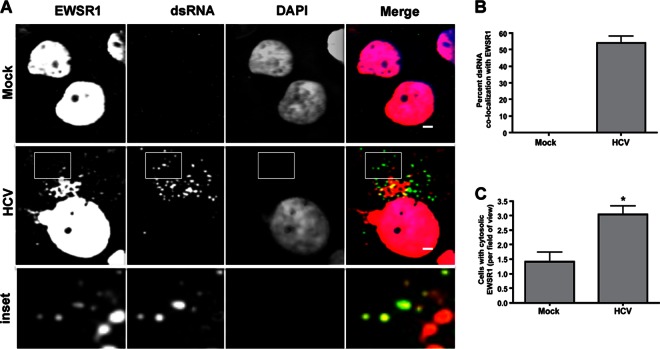
Fig 4.
Localization of HCV replication complexes with EWSR1. (A) Huh-7.5 cells were infected with HCV for 48 h. Cells were fixed and probed with antibodies to EWSR1 (red) and dsRNA (green), the latter detecting HCV replication complexes. DAPI (blue) was used as a nuclear marker. The indicated color is relevant to the merged image. (B) ImageJ quantification of percent cytoplasmic EWSR1 staining localizing with dsRNA replication marker. (C) Quantification of the number of cells positive for cytoplasmic staining in infected and uninfected cell fields of view. Scale, 1 μm. *, P ≤ 0.05.

