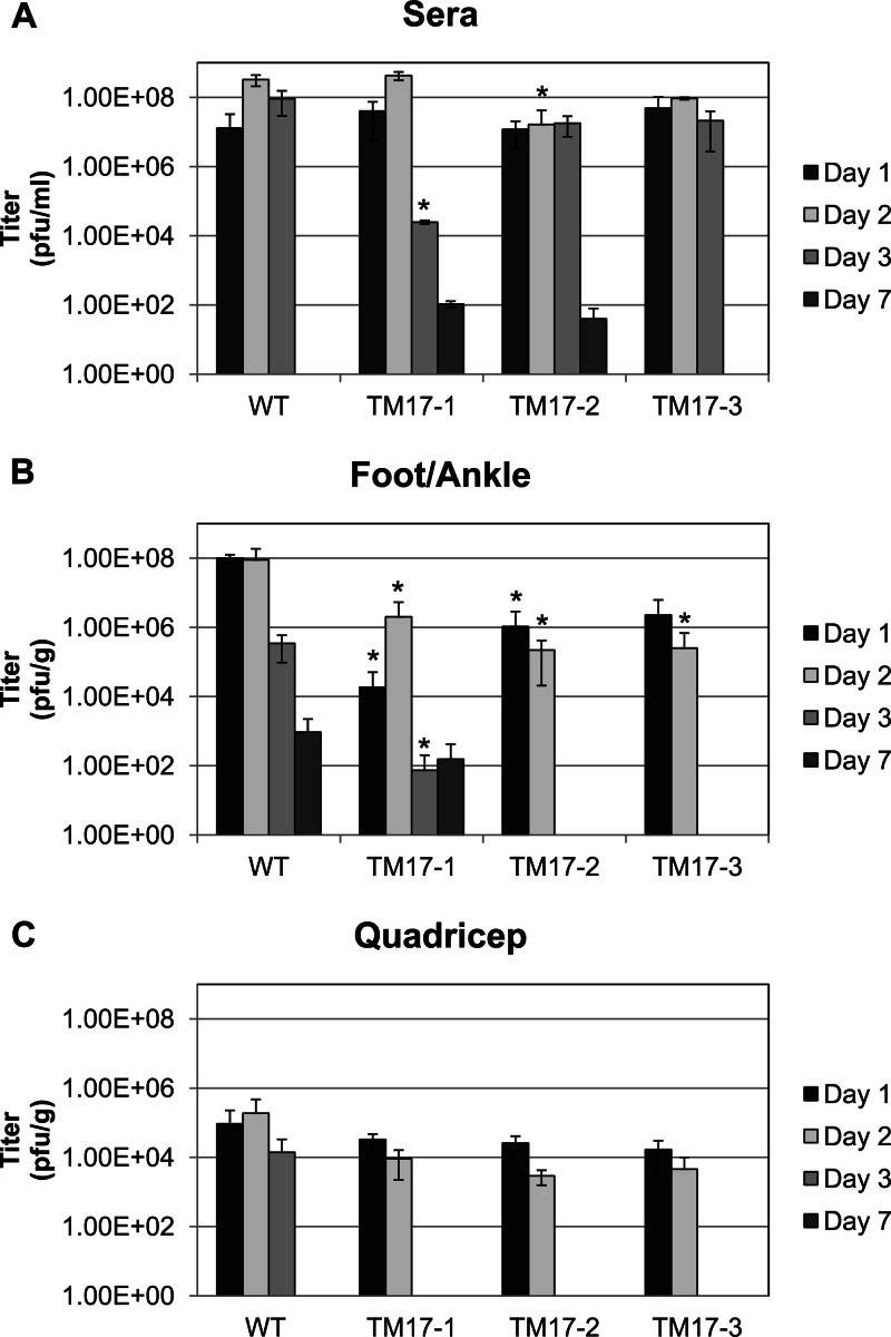
Fig 1.
Prechallenge viremia by plaque assay in mosquito cells in the designated tissues at 1, 2, 3, and 7 days after injection with 103 PFU of WT ChikV, TM17-1, TM17-2, or TM17-3. The values of the mutant virus compared to the WT viremia were analyzed by Welch-corrected Students' t test, and the asterisks indicate where significant differences were found. The error bars indicate standard deviations within sample groups. (A) Viremia detected in mouse sera; analysis of the titers showed no significant difference between the mutants and the WT until day 2 for TM17-2 (P < 0.05) and day 3 (P < 0.001) for TM17-1. (B) Foot and ankle tissue titers differed from WT as follows: day 1, P < 0.001 for TM17-1 and P < 0.01 for TM17-2; on day 2, TM17-1, -2, and -3 titers were significantly lower (P < 0.05). One day 3 virus was cleared from the TM17-2/3-infected mice. However, the WT and TM17-1 were not cleared from mouse feet/ankles at day 7. (C) Titers from quadriceps from which all the mutant viruses were cleared by day 3. No viremia was detected in mice injected with mock samples. The limit of detection of the plaque assay was 40 PFU.

