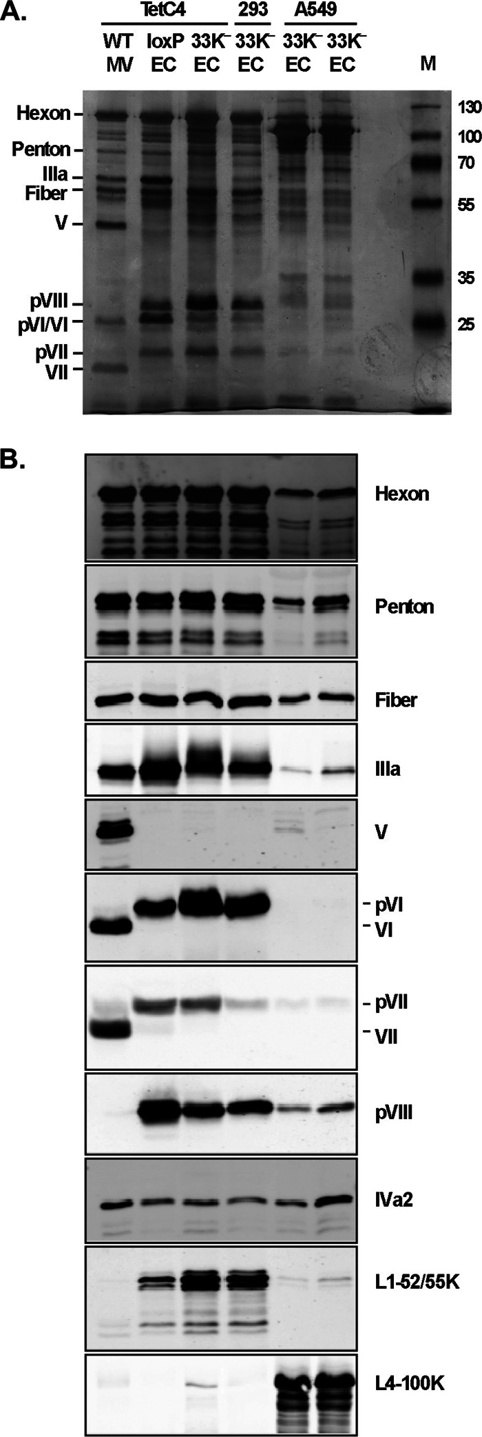
Fig 5.
Protein composition of the empty capsids generated by 33K− virus. (A) Silver staining of protein components of 2 μg of MV and EC isolated from virus particles indicated in Fig. 4A. Protein designations are indicated on the left. M, molecular weight markers. (B) Western blot analysis of protein components of 2 μg of MV and EC isolated from virus particles indicated in panel A. Protein designations are indicated on the right. Virus particles used in these analyses were banded twice by CsCl equilibrium centrifugation. Two independent batches of 33K− EC samples from A549 cells were prepared and included in the analysis (adjacent lanes).

