
Fig 2.
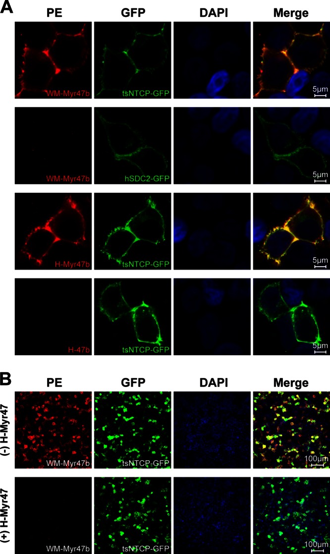
Specific binding of WM-Myr47b peptide to cell surface tsNTCP. (A) 293T cells were transiently transfected with plasmids encoding tsNTCP-GFP or hSDC2-GFP as a control. Transfected cells were cultured in PMM for 36 to 48 h, blocked with 3% bovine serum albumin–phosphate-buffered saline for 1 h, and then incubated with the indicated peptides at 400 nM at 37°C for 2 h. Subsequently, the cells were fixed with 4% paraformaldehyde, stained with 0.6 μg/ml PE-streptavidin, and visualized with a Zeiss LSM 510 Meta confocal microscope. (B) 293T cells transfected with tsNTCP-GFP were blocked with 3% bovine serum albumin–phosphate-buffered saline in the absence (top) or presence (bottom) of 800 nM nonbiotinylated HBV pre-S1 peptide, H-Myr47, followed by washing and incubation with the biotinylated WMHBV pre-S1 peptide, WM-Myr47b, at 400 nM at 37°C for 2 h. Cells were then stained and visualized as described for panel A. DAPI, 4′,6-diamidino-2-phenylindole.