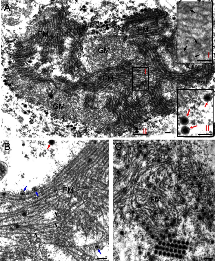
Fig 2.
Transmission electron micrographs of the viroplasms induced by SRBSDV infection in VCMs. (A) The viroplasm matrix consists of granular and filamentous regions. Insets I and II are enlarged images of boxed areas I and II, respectively. (B) Within the filamentous viroplasm matrix, the core of the single-layer particles was densely stained, indicating that viral RNAs are packaged into empty core particles. (C) Within or at the periphery of the filamentous viroplasm matrix, double-layer viral particles accumulated. Black arrows mark single-layer empty particles (∼50 nm in diameter) on the edges of filaments. Blue arrows show single-layer core particles (∼50 nm in diameter). Red arrows mark intact double-layer viral particles (∼70 nm in diameter). GM, granular matrix; FM, filamentous matrix. Bars, 100 nm.

