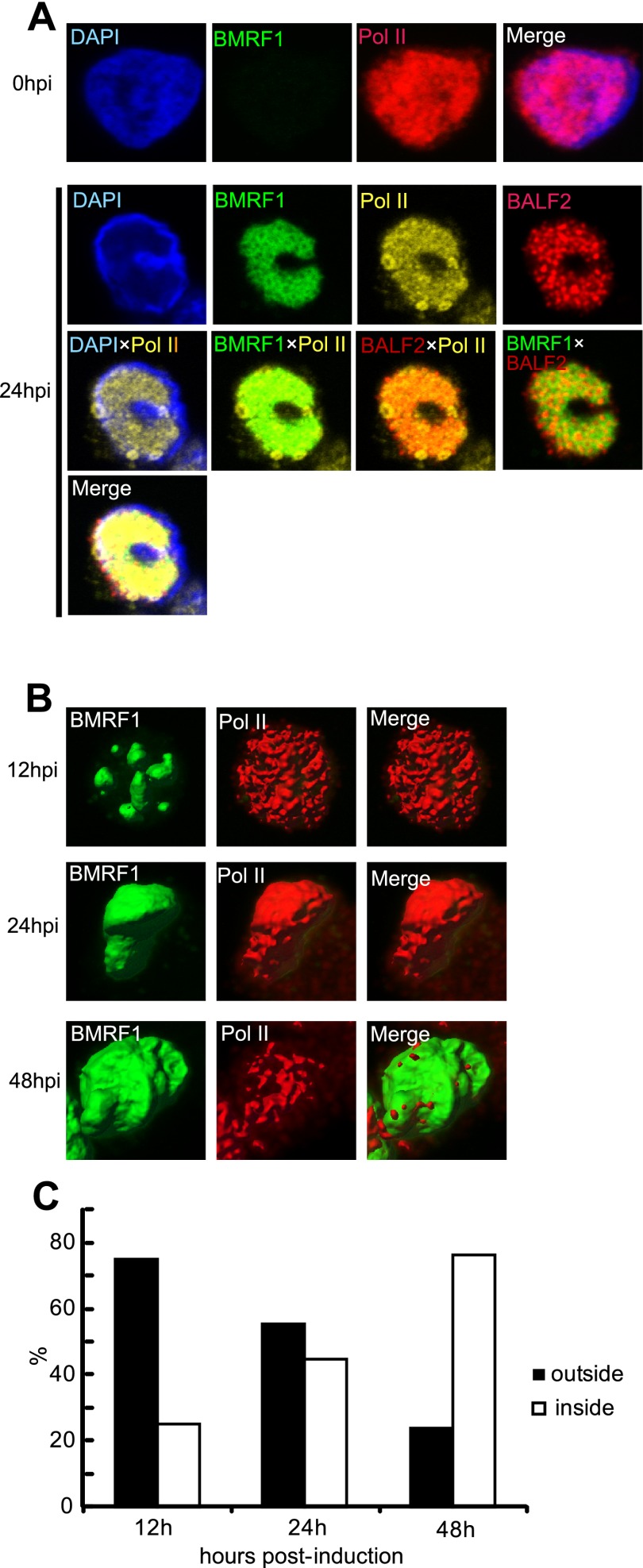
Fig 1.
RNAPII becomes localized inside BMRF1 cores at late stages of productive replication. Tet-BZLF1/B95-8 cells were treated with doxycycline to induce lytic replication and harvested at the indicated postinduction times. After treatment with mCSK buffer, they were fixed. (A) Harvested cells were stained with anti-BMRF1 (green), anti-BALF2 (red), and anti-RNAPII (yellow) antibodies and observed by laser scanning confocal microscopy. The 2D images show brightest-point projections. Lower panels are merged images of the indicated combinations of the proteins. Pol II, RNA polymerase II. (B) Lytic replication-induced cells were stained with anti-BMRF1 (green) and anti-RNAPII (red). Projections of 60 images collected at 0.33-μm steps in the z axis are displayed as 3D topographical reconstruction images of BMRF1 and RNAPII (left and middle panels, respectively). Representative 3D surface reconstruction images are presented. Right panels are merged 3D surface reconstruction images. (C) The ratio of cells in which RNAPII was located outside the BMRF1 core to total BMRF1-positive cells was determined (black bar). White bars show the ratios of cells in which RNAPII was located inside the BMRF core to total BMRF1-positive cells. We regard cells as inside when 50% or more of RNAPII signal was detected inside the core. More than 100 cells were counted at each of the indicated time points.

