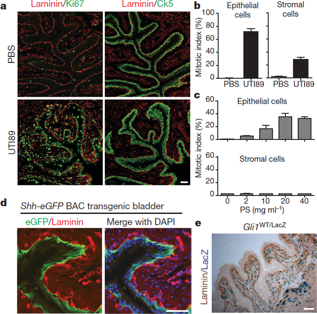
Figure 1. Injury-induced proliferation and Hedgehog signalling in the bladder.
a, UTI89 instillation induces proliferation of basal epithelial and stromal cells of the bladder. Ki67, Ck5 and laminin immunostaining highlight proliferation, basal epithelial cells and the basement membrane, respectively, in bladders 24 h after instillation of UTI89. Adjacent sections were 10 mm apart. b, Quantification of epithelial and stromal cell proliferation in response to bacterial injury. Ki67-positive cells are shown as a per cent of total 4′,6-diamidino-2-phenylindole (DAPI)-staining nuclei. c, Quantification of epithelial and stromal cell proliferation in response to chemical injury. Ki67-positive cells are shown as a per cent of total DAPI-staining nuclei 24 h after instillation of the indicated concentrations of PS. Note the absence of a proliferative response in the stroma. For panels b and c, data are from 3 bladders, 2 sections each, and are shown as mean ± s.e.m.; numerical data are in Supplementary Tables 1 and 2, respectively. d, Expression of eGFP in basal epithelial cells from a Shh-eGFP BAC transgenic mouse. e, Gli1-LacZ expression in the stromal compartment. Bladder sections from Gli1LacZ/WT mice were co-stained with X-gal and anti-laminin. Scale bars in panels a, d and e represent 50 µm.

