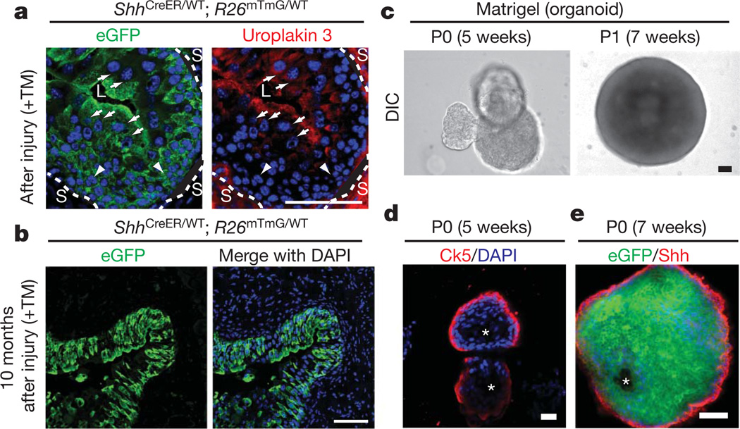
Figure 2. Shh-expressing basal cells repopulate the urothelium and form organoids in vitro.
a, eGFP-marked Shh-expressing cells generate luminal cells positive for uroplakin 3. ShhCreER/WT; R26mTmG/WT mice were treated with TM and subjected to three rounds of injury. Uroplakin-3-positive and negative cells are denoted by white arrows and arrowheads, respectively. Dotted lines demarcate urothelium and stroma (L, lumen; S, stroma). b, Long-term regenerative capacity of eGFP-marked Shh-expressing cells. After TM injection and seven rounds of injury over 10 months, mG expression in bladder from ShhCreER/WT; R26mTmG/WT mice marks most urothelial cells. See Supplementary Fig. 2b for experimental schemes. c, Extended culture of Shh-expressing cells in Matrigel. Single eGFP-positive bladder cells isolated from TM-injected ShhCreER/WT; R26mTmG/WT mice and cultured in Matrigel for 5 weeks formed organoids. Dissociated cells from primary culture (P0) organoids also generated new organoids on subsequent passage (P1) in Matrigel culture. DIC, differential interference contrast. d, Confocal analysis of a bladder organoid. The organoid has multiple layers of epithelial cells with Ck5-expressing cells in the outer layer that contacts the extracellular matrix, and inner cells that line a luminal space and do not express Ck5. e, A section through the wall of an organoid grown in Matrigel and immunostained for Shh. Note eGFP expression in all cells, indicative of Shh expression in the cell initially cultured, but loss of Shh immunostaining from cells that are not in the outer layer. Scale bars represent 50 µm; asterisks denote organoid lumen.

