Figure 3.
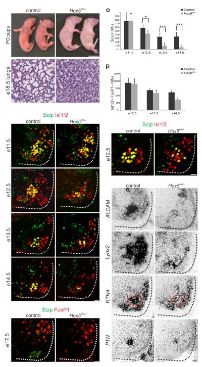
Respiratory failure and PMC loss in Hox5 MNΔ mice. (a–b) All Hox5MNΔ mice are born cyanotic and perish at birth. (c–d) Histological analysis reveals that the lungs of e18.5 Hox5MNΔ embryos collapse before birth. (e–n) Progressive loss of Scip+ PMC neurons in Hox5MNΔ mice. At e11.5 Scip+ neuron numbers are similar between control and Hox5MNΔ mice but progressively decrease in the mutants. By e17.5 there are no detectable Scip+ motor neurons in Hox5MN mice. (o) Quantification of Scip+ motor neurons in control and Hox5MNΔ mice. At least 5 pairs of embryos were analyzed for each time point. *P<0.05, ***P<0.001 (p) Quantification of non-LMC Isl1/2+ motor neurons. There is selective loss of FoxP1− neurons in Hox5MNΔ mice. (q–r) PMC disorganization at e12.5 in Hox5MN mice. Isl1/2+ Scip– neurons are intercalated with PMC neurons. (s–t) ALCAM expression is reduced in Hox5MN mice, as early as e11.5 prior to loss of Scip. (u–z) Lynx2, RTN4 and PTN are downregulated in Hox5MN mice at e12.5. PMC position is outlined by dashed red line. Expression of Lynx2 is also lost from LMC neurons. Scale bars=25μm, except in (b)=1cm and (d)=100μm. Error bars represent s.e.m.

