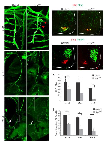
Figure 4.
Fidelity of PMC axon projections in Hox5MNΔ mutants. (a–f) The phrenic nerve progressively thins in Hox5MNΔ mutants. At e12.5 phrenic nerve diameter is similar between control and Hox5MNΔ mice (a–b). Some axons stray from the phrenic nerve in Hox5MNΔmice (arrows in b). By e14.5 the phrenic nerves become thinner in Hox5MNΔmice (see inserts e–f) and lack arbors seen in control nerves (arrows in e–f). (g–j) Retrogradely labeled motor neurons after RhD injection in the phrenic nerve are reduced in Hox5MNΔ mice, but retain some aspects of their molecular identity, such as Scip expression (g–h) and FoxP1 exclusion (i–j). (k–l) Quantification of RhD retrograde transport after RhD injection in the phrenic nerve. There is a decrease in the number of RhD+ neurons in Hox5MNΔ mice (k), as well as a decrease in the percentage of RhD+ neurons that express Scip (l). Scale bars=100μm (a–f), 25 μm (g–j). Error bars represent s.e.m., *P<0.05,**P<0.01.

