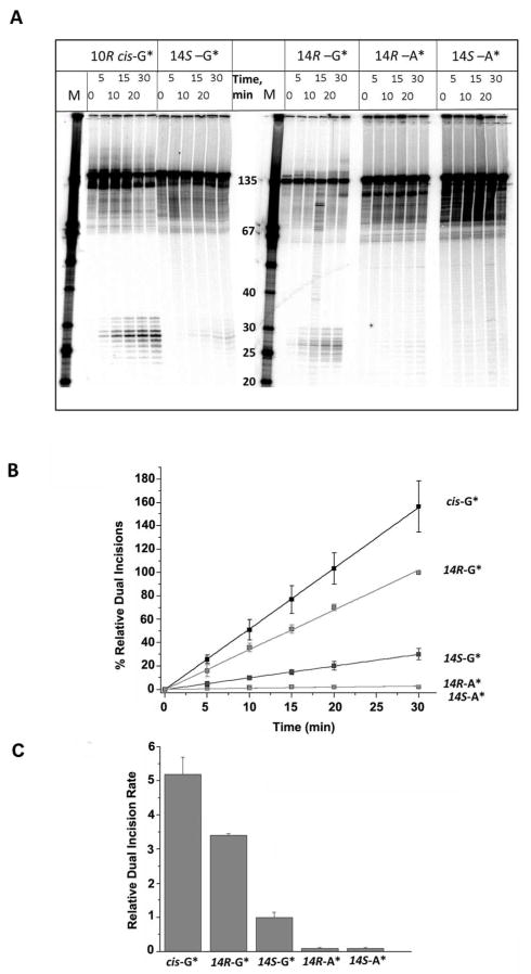
Figure 2.
(A) Autoradiogram of denaturing gels showing the appearance of NER dual incision products (~ 24 – 32 nucleotides in length) after incubating 135-mer duplexes with single DNA adducts in HeLa cell extracts for the time intervals indicated. The lanes containing unmodified single-stranded oligonucleotide size markers with 3′- and 5′-OH groups at the ends and the indicated number of nucleotides in lengths are shown in the lanes marked M. The results shown were all obtained with aliquots from the same incubated sample but the gel electrophoresis was conducted in two different gels (two samples on the left, and three samples on the right, respectively). The absolute % excision in this example was 8.7% in the case of the 14R-DB[a,l]P-N2-dG adduct (30 min time point), but varied from 6 – 17% in the other four cell extracts. Other details are discussed in Supporting Information. The densitometry tracings of some of the lanes are shown in Supporting Information, Figure S4. (B) Time course of NER dual incision product formation of 24 – 32 oligonucleotide fragments bearing either the R-cis-B[a]P-N2-dG (cis-G*), R-DB[a,l]P-N2-dG (14R-G*), S DB[a,l]P-N2-dG (14S-G*), R-DB[a,l]P-N6-dA (14R-A*), or the S-DB[a,l]P-N6-dA (14S-A*) adducts. The experimental data points are averages of five independent experiments in different cell extracts, and the error bars represent the standard deviations. (C) Comparisons of initial rates of dual incisions derived from the linear plots in panel B.

