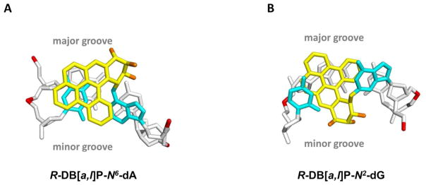
Figure 5.

(A) View looking down the helix axis of the (A6*:T17)•(C7:G16) segment in the R-DB[a,l]P-N6-dA adduct. (B) View looking down the helix axis of the (G6*:C17)•(C7:G16) segment in the R-DB[a,l]P-N2-dG adduct. The view is in the 5′ to 3′ direction along the lesion-containing strand. Stereo views of (A) and (B) are provided in Supporting Information Figure S7.
