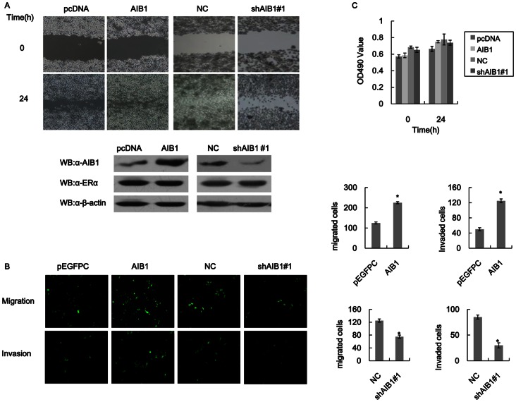
Figure 2. AIB1 promotes cell motility and invasion in T47D cells.
(A) Scratch wound-healing assay showing the effect of AIB1 on cell motility in T47D cells. Top panel: Images of T47D cells that were transfected with empty vector (pcDNA) or AIB1, and cells without (NC) or with AIB1 knockdown (shAIB1#1) before and after wound-healing assay. The cell layers were carefully wounded using a sterile 200-µl tip and then cultured for 24 h before evaluation. Bottom panel: Western blot analysis showing the levels of AIB1, ERα and β-actin expressions in pcDNA- or AIB1-transfected T47D cells, and of T47D cells without or with AIB1 knockdown. (B) Transwell migration and invasion assays showing the effect of AIB1 on cell motility and invasion ability in T47D cells. Images showing the migration and invasion of T47D cells that were transfected with empty vector (pcDNA) or AIB1, and of T47D cells without (NC) or with AIB1 knockdown (shAIB1#1). For NC group, the cells were transfected with a negative control scrambled shRNA synthesis DNA cloned into siRNA expression vector pRNAT carries GFP marker. Cell migration and invasion assays were performed in 24-well chambers without and with Matrigel, respectively. Cells (1000 per well) were transfected with GFP-AIB1 or just GFP (pEGFPC) and then transferred to the upper chamber. After 48 h of incubation, the numbers of migrating and invasive cells on the lower surface of the filter were counted under a fluorescent microscope. The bar graphs on the right of the images show the number of migrating and invading cells for each category of cells. (C) MTT assays showing the effect of AIB1 on cell proliferation in T47D cells. Cells were transfected as in (A) and then subjected to MTT assay within 24 hours.

