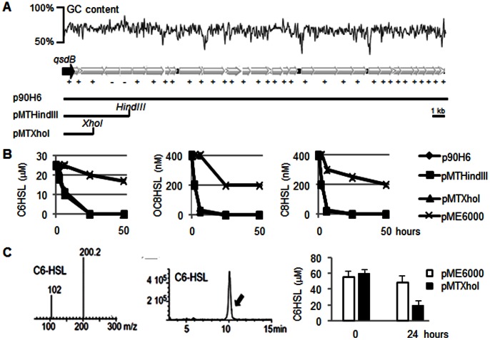
Figure 4. Physical map of the fosmid p90H6 and sub-cloning of qsdB coding for NAHLase activity.
In A, GC content (%) and orientation (+/−) of the 34 putative orfs of the fosmid p90H6, and physical map of the pME6000-derivatives pMTHindIII and pMTXhoI barboring qsdB (orf1). In B, residual level of C6HSL, OC8HSL or C8HSL measured in the presence of E. coli strain DH5α harboring the empty vector pME6000, the fosmid p90H6, and the constructed pMTHindIII and pMTXhoI (symbols of the three formers are superimposed in the graphs).Three replicates were done. In C, C6HSL analysis by HPLC/MS: mass, retention time, and quantification of C6HSL before (t = 0) and 24 hours after incubation in the presence of cell-free extracts of E. coli strains DH5α(pME6000) and DH5α (pMTXhoI).

