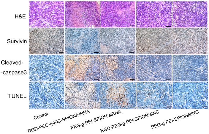
Figure 7. Ex vivo histological analyses of tumor tissue sections.
The H&E staining, immunohistochemical and TUNEL analyses of tumor tissue sections from the mice treated with various complexes formed at a N/P ratio of 10. In the immunohistochemical assay, the brown stains indicate Survivin or cleaved caspase-3 protein. In the TUNEL assay, the brown stains indicate apoptotic cells (×200; scale bar:100 µm; Control: the mice injected with PBS).

