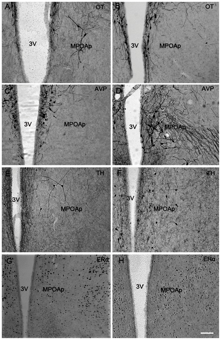
Figure 7. Photomicrographs of neuropeptides expression in the posterior subnucleus of the medial preoptic area (MPOAp).
Photomicrographs displaying OT-ir (A & B), AVP-ir (C & D), TH-ir (E & F) and ERα-ir (G & H) stained cells in the posterior subnucleus of the medial preoptic area (MPOAp) in the brains of Mongolian gerbils (A, C, E & G) and Chinese striped hamsters (B, D, F & H). 3V, third ventricle, Scale bar = 100 µm.

