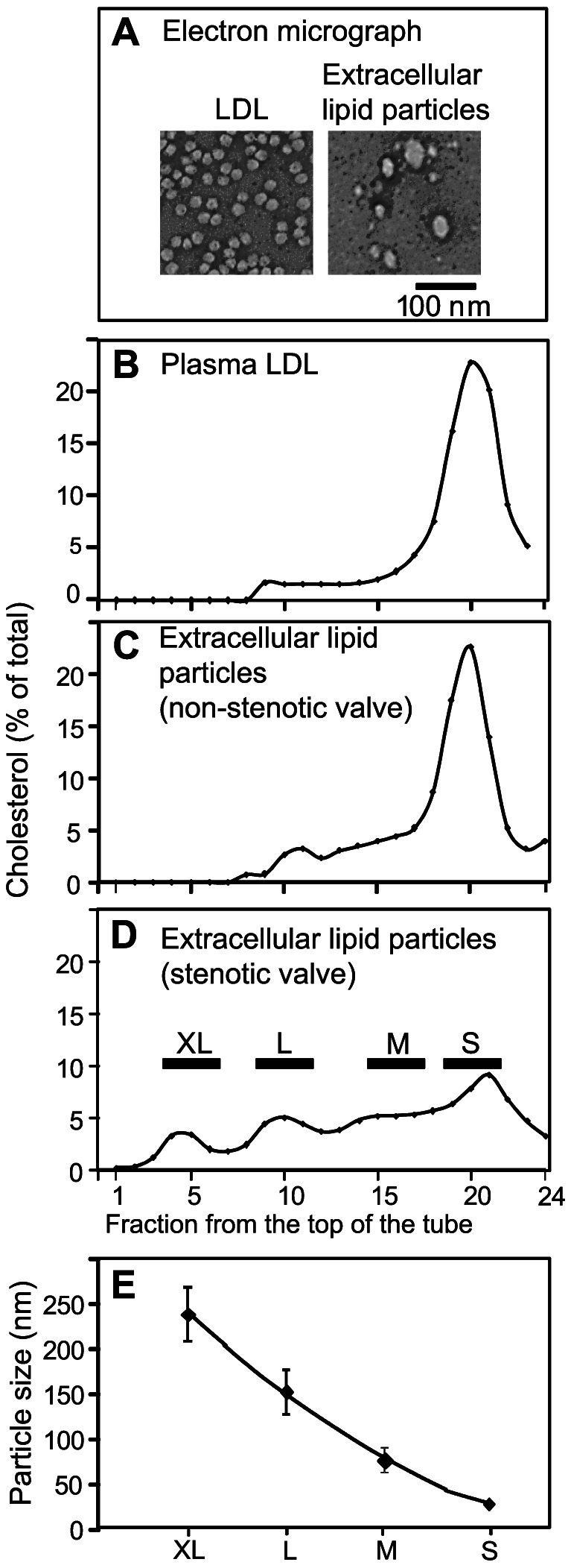
Figure 3. Electron micrographs and rate zonal ultracentrifugation of extracellular lipid particles.

The extracellular lipid particles and native LDL were negatively stained and photographed under electron microscope as described in the Methods (A). Native LDL (B), lipid particles from a non-stenotic valve (C), and lipid particles from a stenotic valve (D) were subjected to rate zonal ultracentrifugation as described under Methods. Fractions (500 µl) were collected and their cholesterol concentrations were determined. The fractions in each sample were pooled into four groups based on the floating pattern of the extracellular lipid particles of the stenotic valves (D). The particle sizes in each pool were determined using dynamic light scattering and the average sizes of each particle class are shown in Panel E.
