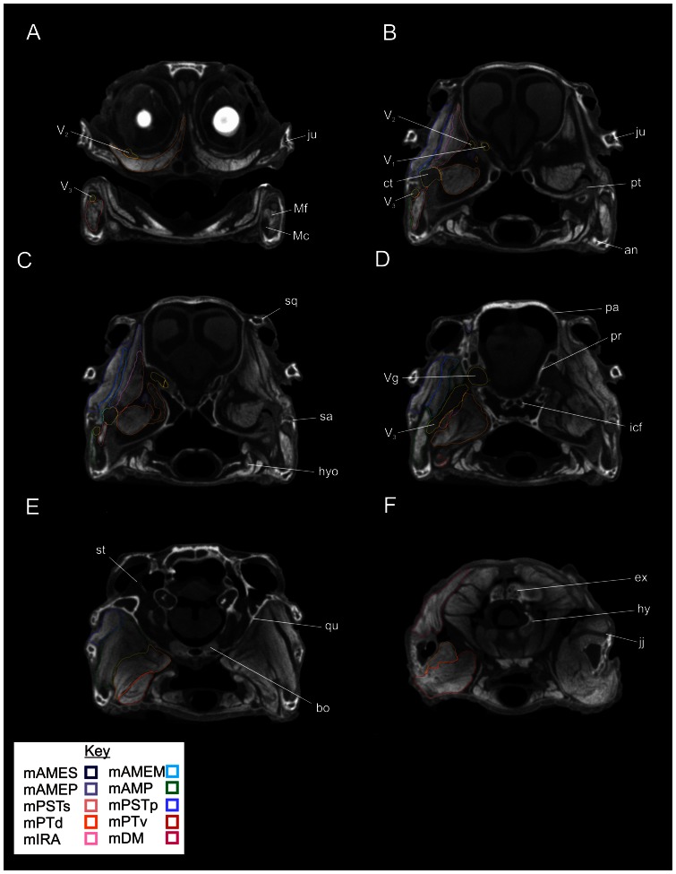
Figure 5. Rostral views of axial sections through the segmented CT data showing labeled individual right jaw muscles on the left, and unlabeled left structures on the right.
A, Slice 391/911; B, Slice 316/911; C, Slice 305/911; D, Slice 283/911; E, Slice 240/911; F, Slice 154/911. Abbreviations: an, angular; bo, basioccipital; ct, cartilage transiliens; ex, exoccipital; hyo, hyoid; icf, internal carotid foramen; jj, jaw joint; ju, jugal; Mc, Meckel's cartilage; Mf, Meckelian fossa; naa, neural arch of atlas; pa, parietal; pr, prootic; pt, pterygoid; qu, quadrate; sa, surangular; sq, squamosal; st, stapes; V1, ophthalmic nerve; V2, maxillary nerve; V3, mandibular nerve; Vg, trigeminal ganglion.

