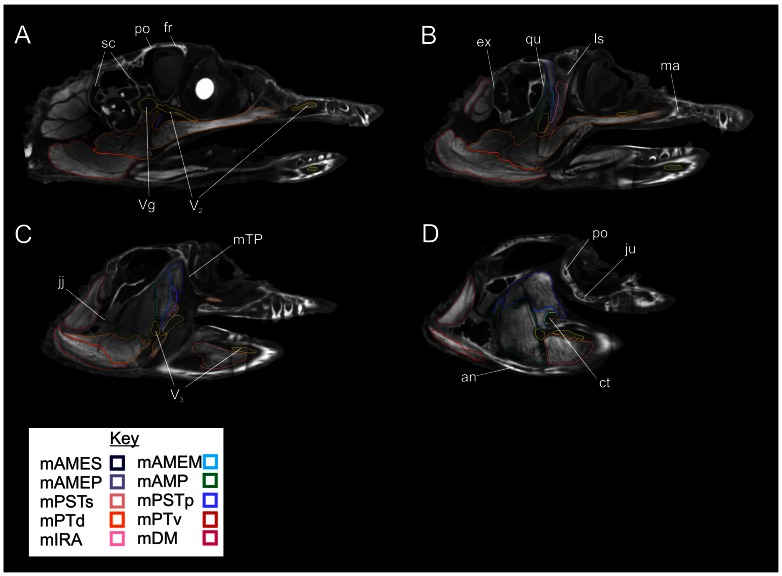
Figure 6. Lateral views of parasagittal sections through the segmented CT data showing labeled individual right jaw muscles.
A, Slice 183/512; B, Slice 155/512; C, Slice 133/512; D, Slice 119/512. Abbreviations: an, angular; ct, cartilago transiliens; ex, exoccipital; ju, jugal; ls, laterosphenoid; ma, maxilla; mTP, m. tensor periorbitae; po, postorbital; qu, quadrate; sc, semicircular canals; V2, maxillary nerve; V3, mandibular nerve; Vg, trigeminal ganglion.

