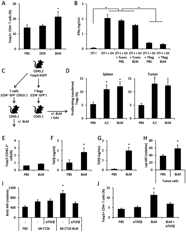
Figure 4. BLM induces in vivo Treg accumulation throught TGFb production.
A: Spleens from CT26 tumor-bearing mice, treated with PBS or BLM, were harvested and the cells were analyzed through flow cytometry for Foxp3+CD4+ Treg detection. B: Treg from PBS- or BLM- treated CT26-bearing mice were isolated from spleen and co-cultured with OT-I in presence of SIINFEKL (SII). After 3 days of culture, IFNγ was titrated in the supernatant using ELISA method. C: Schematic representation for D and E panel experiments. D: GFP+ CD4+ cells were sorted from CD45.2 FOXP3-EGFP mice, then injected i.v. in CD45.1 mice bearing CT26 tumor. The mice then received PBS, IL-2 or BLM treatment. All mice received EdU injection. One day after treatment, spleens and tumors were collected and the proliferation status of transferred cells was assessed by revealing EdU by flow cytometry. E: CD4+ CD62L+ GFP− naive T cells were sorted from CD45.2 FOXP3-EGFP mice, and injected i.v. in CD45.1 mice bearing CT26 tumor. The mice then received PBS or BLM treatment. Spleens were harvested and the cells were analyzed through flow cytometry for Treg detection. F: Mice were injected with 1.106 CT26 cells i.p. Ten days later mice received PBS or BLM injection. The following day ascites were collected, and TGFβ was assessed using ELISA method. G: CT26 cells were treated with PBS or BLM in vitro for 24 h, then the supernatant was collected and assessed for TGFβ using ELISA method. H: Mice bearing CT26 tumor cells were treated with PBS or BLM. The day after, CT26 tumors were collected and tumor cells were separated from the tumor infiltrating lymphocytes. Tumor cells were stained for LAP and analyzed by flow cytometry. I: GFP+ CD4+ cells were sorted from CD45.2 FOXP3-EGFP mice, then cultured in vitro under TCR-stimulating conditions. We added culture supernatant of CT26 treated for 24 h with PBS of BLM. In some wells, blocking anti-TGFβ antibody was added. After three days of culture, the cells were incubated with BrdU for 3 h, and then proceeded to BrdU detection by flow cytometry. J: Same as A, and mice received injection of blocking anti-TGFβ antibody. All data presented are representative of one out of two (panels B, D, E, G and I) or three experiments (panels A, F, H and J). Graphs show mean +/− SEM.

