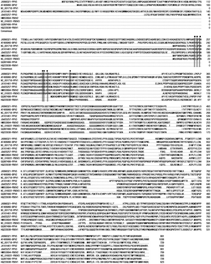
Figure 2. Amino acid sequence alignment of VP1 with the putative phospholipase A2 motif of PPV5, other PPVs, PBoV, and BPV2.
The Ca2+ binding loop is indicated by filled squares and the catalytic residues are indicated by filled circles. “.” indicates a deletion compared to the strain on top. The positions of the amino acids and the GenBank numbers of the sequences are indicated.

