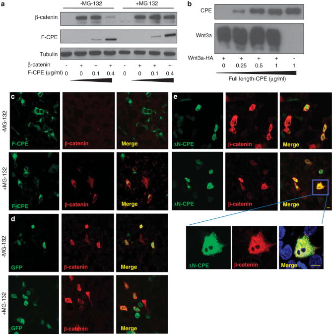
Figure 3.
The effect of CPE on the Wnt signal depends on the proteasome activity. (a) HEK293T cells were co-transfected with GFP-β-catenin and F-CPE-GFP as indicated. Twenty-four hours later, 25μm MG132 was added to the cells for an additional 24 h. Cells were harvested and subjected to western blot analysis using an anti-GFP antibody. Tubulin served as a loading control. (b) HEK293T cells were co-transfected with Wnt3a-HA and increasing amounts of F-CPE-GFP as indicated. Western blot analysis was performed using anti-HA and anti-GFP antibodies, respectively. (c, d and e) HEK293T cells were co-transfected with F-CPE-GFP, GFP or the ΔN-CPE-GFP constructs along with myc-β-catenin (as indicated). Twenty-four hours post transfection the cells were treated overnight with 25 μm MG132. The cells were fixed and reacted with an anti-myc antibody and rhodamine-conjugated antibody. Images were taken using a confocal microscope. Note the ΔN-CPE immunostaining in the cytoplasm and the nucleus in some cells. Bar = 10μm.

