Figure 4.
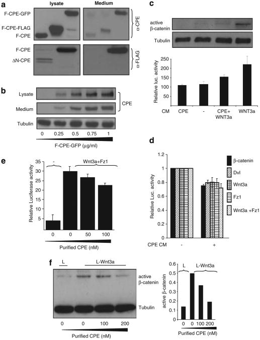
Secreted CPE affects Wnt signaling. (a; upper panel) HEK293T cells were transfected with F-CPE-GFP, F-CPE-FLAG and untagged-F-CPE. Forty-eight hours post transfection the cell media was collected and the transfected cells were harvested. The media and lysates were subjected to western blot analysis using an anti-CPE antibody. HEK293T cells were transfected with FLAG-tagged F-CPE and ΔN-CPE (lower panel). Forty-eight hours post transfection the cell media was collected and the transfected cells were harvested. The media and lysates were subjected to western blot analysis using an anti-FALG antibody. Tubulin served as a loading control. (b) HEK293T cells were transfected with different amounts of F-CPE-GFP. Media and lysates were subjected to western blot analysis using an anti-CPE antibody. (c) HEK293T cells were transfected with F-CPE-GFP. Twenty-four hours later, CM was collected from transfected cells. Wnt CM was collected from L-Wnt3a cells. The different media was added to HEK293T cells transfected with the pTOPFLASH/pFOPFLASH reporter plasmids. Luciferase levels were detected 24 h later. The upper panel shows the levels of endogenous, active β-catenin as detected using a specific antibody. (d) HEK293T cells were co-transfected with β-catenin-GFP, HA-Wnt3a, HA-Dvl and Fz1 along with the pTOPFLASH/pFOPFLASH reporter plasmids. CPE CM (as in c) was added to transfected cells for 24 h and luciferase levels were then detected. (e) HEK293T cells were co-transfected with Wnt3a and Fz1 along with the pTOPFLASH/pFOPFLASH reporter plasmids. Twelve hours later the cells were supplemented with media containing purified CPE as indicated. Twelve hours later luciferase levels were determined. (f) L and L-Wnt3a cells were supplemented with media containing purified CPE as indicated. The cells were harvested 12 h later and active β-catenin levels were detected using a specific antibody. Tubulin served as a loading control (left). Densitometric analysis of active β-catenin was performed using TINA software (right).

