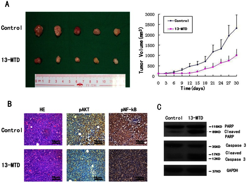
Figure 5.
The therapeutic effect of 13-MTD on Jurkat cell xenografts.
The tumor volumes of xenografts were measured with calipers every 3 days for a total of 30 days after the start of treatment. After 30 days of treatment, mice were sacrificed and the tumors removed and photographed. (A) Tumor growth was significantly suppressed with 13-MTD treatment. The respective tumor volumes from the solvent control and 13-MTD treatment groups were 2325.43±318.32 mm3 and 1000.54±156.78 mm3 (n = 5, P<0.01, Student’s t-test) (B) Tumors were then fixed and stained with H&E to examine tumor cell morphology. IHC showed decreased phosphorylation of AKT and NF-κB in the tumor tissue after treatment with 13-MTD. (Original magnification×100) (C) 13-MTD enhanced the activation of caspase-3 and PARP proteins in tumor xenografts compared with controls. Tumor lysates were subjected to the analysis of protein levels using western blot analysis. GAPDH was used as a loading control. Representative blots are shown from independent experiments with six different tumors in each treatment group.

