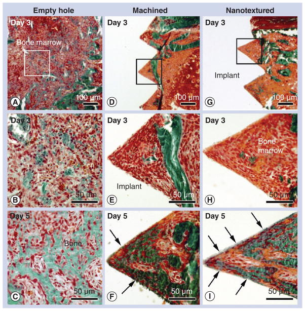
Figure 3. Histology of bone formation in an empty hole and around machined and nanotextured implants.
Light microscope images of calcified sections stained with Goldner-trichrome of the tissue (A, B & C) formed in an empty hole, and (D, E & F) around machined and (G, H & I) nanotextured screw-shaped implants at (A, B, D, E, G & H) day 3 and (C, F & I) 5 post-surgery. With time, there is an increase in bone formation in the marrow at the surgical site left empty (empty hole) as well as around either type of implant. At day 3, bone formation was hardly noted around both types of implants. At 5 days postimplantation, bone–implant contact (arrows) is more advanced around nanotextured implants (I) than machined implants (F). (B, E & H) High-magnification of the boxed areas in (A), (D) and (G), respectively.

