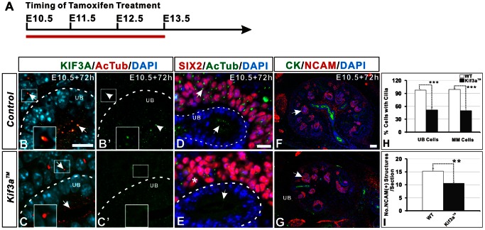
Figure 1. Loss of primary cilia and decreased nephron number in mice with Tamoxifen-induced Kif3a deficiency.
(A) Chart showing embryonic stage at which Tamoxifen was injected (E10.5) and at which kidneys were retrieved (E13.5) for analysis (red line). (B, B’) KIF3A co-localizes with α-AcT in both ureteric bud and metanephric mesenchyme cells in WT kidney at E13.5. Insert box shows high-resolution image of KIF3A located in a primary cilium in a metanephric mesenchyme cell. (C, C’) Expression of Kif3a is largely undetectable 72 hours after Tamoxifen administration to pregnant Kif3aloxP/loxP mice which had been crossed to Cre-ER™;Kif3a+/− mice. (D, E) The number of primary cilia (green) is markedly decreased in both SIX2-positive cells (nephrogenic precursors) and in ureteric cells Kif3a™ kidney (E, asterisk) compared to WT kidney (D). (F, G) Imaging of NCAM-positive nephrogenic precursor structures (arrows). The number of precursors is decreased in Kif3a™ mice (G) compared to WT mice (F). (H) Quantitation of the number of cells with a primary cilium. Tamoxifen administration to Kif3a™ embryos decreases cilia number by approximately 50% in both ureteric and metanephric mesenchyme cells (***, P<0.001). (I) Quantitation of the number of NCAM-positive nephrogenic precursor structures in tissue sections reveals a significant decrease in Kif3a™ mice compared with WT mice (**, P<0.01). WT, wild type; Scale bar: 25 micrometer.

