
Figure 5. EFhd2’s coiled-coil domain mediates the association with tau proteins.
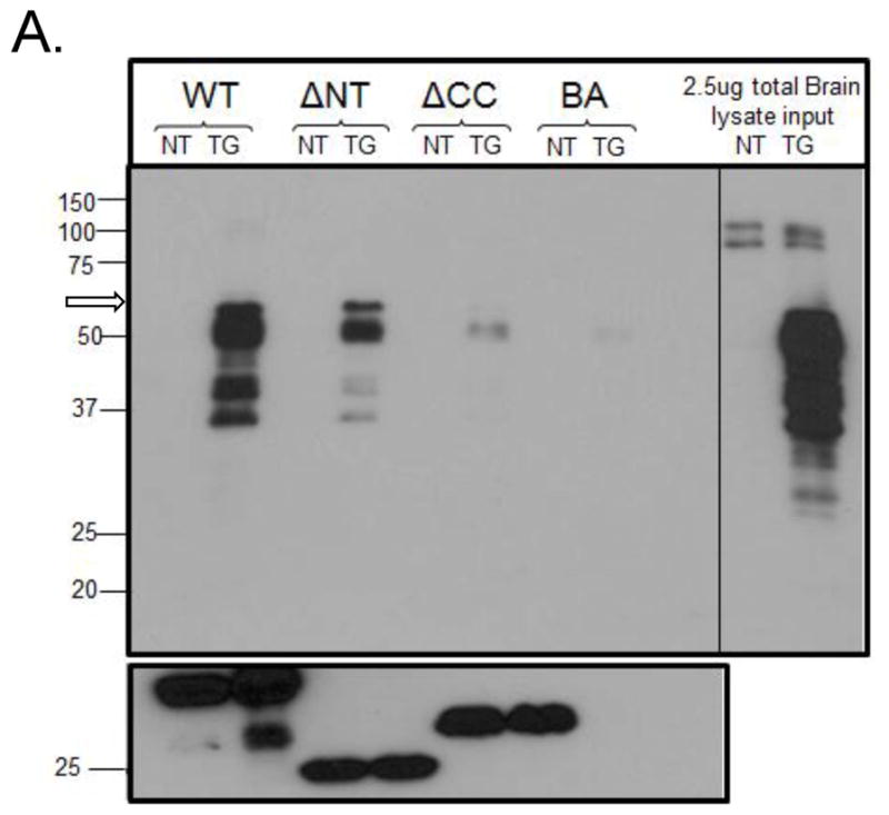
Recombinant His EFhd2WT (WT), His EFhd2 ΔNT (NT) and His EFhd2 ΔCC (ΔCC) proteins were incubated with 11month-old P301L (TG) or an age-matched non-transgenic mouse (NT) whole brain lysate. A control incubated without recombinant proteins was also added (BA). After incubation, proteins were pulled down by their His tag using a nickel column, including the negative control (BA). Proteins bound to the columns were resolved on an SDS-PAGE and an immunoblot against tau (using anti-Tau13) and His (anti-His) was performed. 2.5μg of total protein from TG and NT brain lysates (representing 0.25% total protein input in this experiment) is shown in the right panel. An open arrow (left panel, over 50kDa band) points the migration of the 64kDa band representing hyperphosphorylated tau proteins.