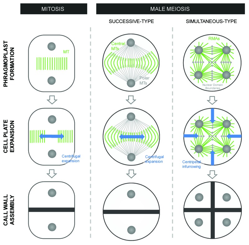
Figure 2. Microtubule array formation and direction of cell wall formation in “conventional” cytokinesis and in male meiosis I and II of respectively successive and simultaneous type PMC cytokinesis. The newly formed cell plate and associated deposition of transient callose is presented in blue. Phragmoplast and RMA microtubule structures are indicated in green and polar MT bundles are shown in gray.

An official website of the United States government
Here's how you know
Official websites use .gov
A
.gov website belongs to an official
government organization in the United States.
Secure .gov websites use HTTPS
A lock (
) or https:// means you've safely
connected to the .gov website. Share sensitive
information only on official, secure websites.
