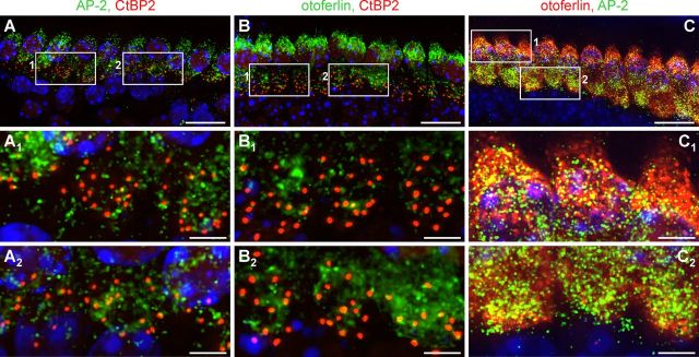
Figure 3.
Comparison of the protein localization of AP-2 and otoferlin with the ribbon marker CtBP2. A–C, Whole-mount preparations of mice IHCs (P18) coimmunolabeled with AP-2 and CtBP2 (A), otoferlin and CtBP2 (B), or otoferlin and AP-2 (C). AP-2 (A1, A2) and otoferlin (B1, B2) showed only minor colocalization with CtBP2 at the basal pole of IHCs as demonstrated in two examples each. In contrast, otoferlin and AP-2 largely colocalized in the apical (C1) and in the basal pole (C2) of IHCs. Cell nuclei were counterstained with DAPI. Scale bars: (in A–C) 20 μm; (in A1,A2-C1,C2) 5 μm.

