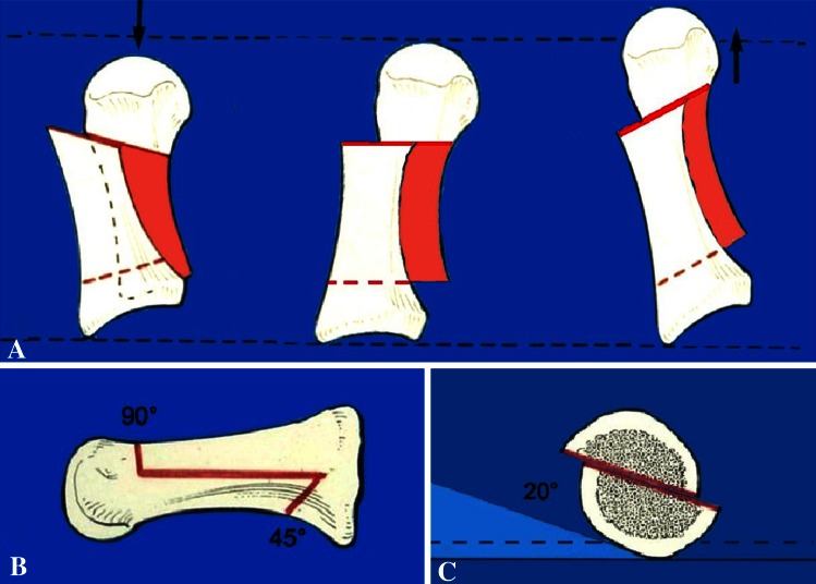
Fig. 5A–C.
The drawings show the Scarf osteotomy technique. (A) The dorsoplantar view shows how to maintain or modify the length of first metatarsal bone. (B) The lateral view shows how to perform a correct osteotomy, and (C) the AP view shows how to correct a first ray insufficiency. The osteotomy must be performed with a 20°-angle and the distal portion is displaced plantarward.

