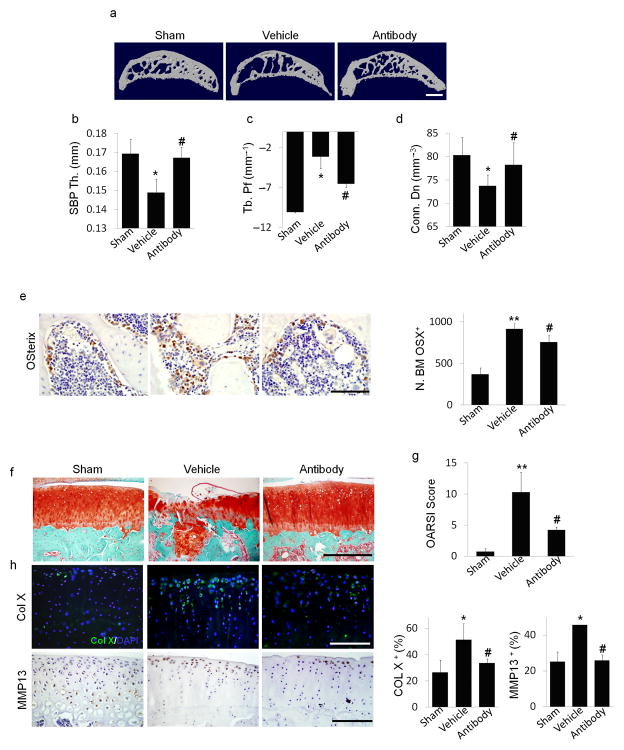
Figure 5. Local subchondral administration of TGF–β antibody reduced abberant subchondral bone formation and articular cartilage degeneration in ACLT rats.
(a) Three dimensional μCT images of tibia subchondral bone medial compartment (sagittal view) in rats that underwent sham (Sham) or ACLT surgery with implantation of an alginate bead containing either vehicle (Vehicle) or TGF–β antibody (Antibody) 3 months post surgery. Scale bar, 1 mm. (b–d) Quantitative analysis of structural parameters of subchondral bone by μCT analysis: thickness of subchondral bone plate (SBP), trabecular pattern factor (Tb. Pf) and connectivity density (Conn. Dn). (e) Immunohistochemical and quantitative analysis of osterix (brown). Scale bars, 100 μm. (f) Sanfranin O–fast green staining of sagittal sections of subchondral tibia medial compartment, scale bar, 400 μm. (g) OARSI scores. (h) Immunofluorescent or immunohistochemical and quantitative analysis of type X collagen (green,) and MMP13 (brown) in articular cartilage. DAPI stains nuclei (blue) (center). Scale bars, 200 μm. n = 8; *P < 0.05, **P < 0.01 vs. sham, #P < 0.05 vs. vehicle ACLT rats.

