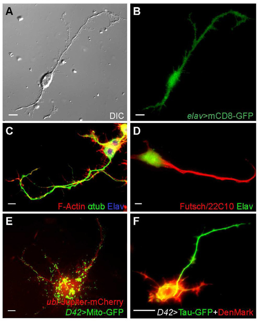
Figure 1. Characterization of cultured Drosophila neurons.
(A–B) A neuron expressing UAS-mCD8-GFP under control of elav-Gal4.
(C–D) Wild-type neurons fixed and stained with TRITC-conjugated Phalloidin, anti-tubulin antibody and anti-Elav antibody (C), or anti-Futsch (22C10) and anti-Elav antibodies (D).
(E) A neuron expressing UAS-Mito-GFP under control of D42-Gal4, and mCherry-Jupiter under ubi promoter.
(F) A neurons expressing UAS-Tau-GFP and UAS-DenMark under control of D42-Gal4.
Scale bars, 5 µm.
See also Movie S1.

