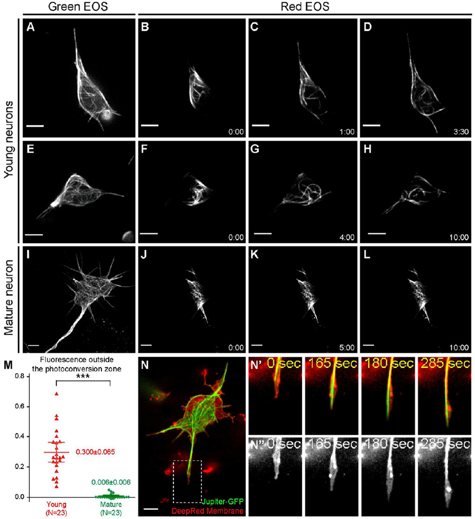
Figure 3. Microtubule sliding drives neurite outgrowth in young neurons.
(A–L) Cultured neurons expressing photoconvertible tdEOS-αtub under maternal αtub-Gal4 and zygotic D42-Gal4. (A,E,I) tdEOS-αtub imaged in the green channel before photoconversion. (B–D,F–H,J–L) tdEOS-atub imaged in the red channel after photoconversion. Time after conversion (in min:sec) is shown in individual frames. Top two rows, young neurons; the third row, mature neuron.
(M) Quantifications of microtubule sliding. Fluorescent intensity outside the photoconversion zone was measured in the red channel 10 min after conversion. 95% confidence interval (CI) for the mean: young neuron=0.300±0.065 (n=23; SEM=0.031; SD=0.150); mature neuron= 0.006±0.006 (n=23; SEM=0.003; SD=0.015). Unpaired t-test between young and mature neurons gives p<0.0001 (***).
(N–N”) A cultured young neuron expressing GFP-tagged endogenous Jupiter (labels microtubules) was stained with DeepRed (cell membrane). The whole neuron (N), and a fast-growing neurite (the dashed box in N): merged channel in top panels (N’) and DeepRed channel in bottom panels (N”). See also Movie S5. Scale bars, 5 µm.

