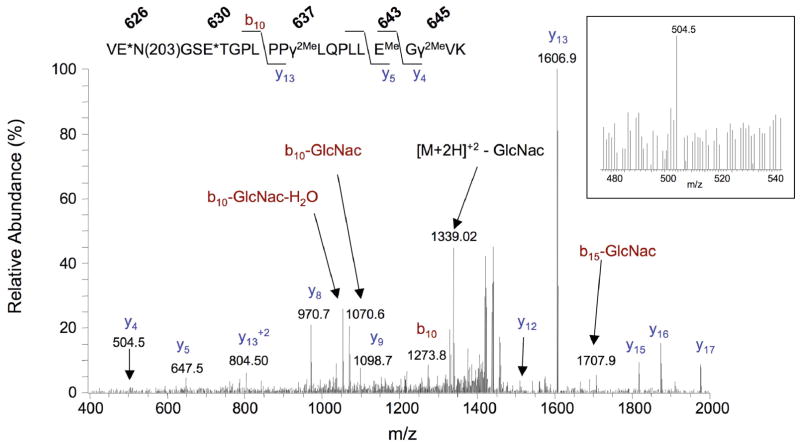
Figure 7. CID spectrum of a Gla-containing carboxylase peptide.
A 625-647 peptide with a mass (2879.3 Da) indicating 3 Gla residues was identified after in-gel trypsin digestion of carboxylase, followed by methylation and redigestion with trypsin (Table S9). EMe, γ2Me and E* denote methylated Glu, dimethylated Gla and either methylated Glu or Gla, respectively. The spectrum shown is for the [M+2H]+2 peptide form. The spectrum for the [M+2H]+3 form showed a higher signal for the y4 ion, which is shown in the boxed insert.

