
Figure 3.
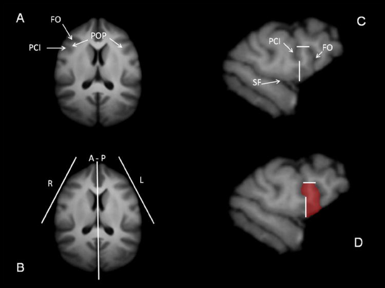
(A) Location of the FO and PCI sulci in relation to the curvature of the frontal cortex as seen in an axial view of the chimpanzee brain template. (B) Orientation of the para-sagittal planes of cutting for the right (R) and left (L) POP. (C) Example of a para-sagittal slice showing the borders defining the POP region. (D) Manual tracing of the POP on the same para-sagittal slice. FO = Fronto-Orbital Sulcus; PCI = Pre-Central Inferior Sulcus; POP = Pars Opercularis; A–P = Anterior-Posterior Axis; SF = Sylvian Fissure